Electron microscopes are very powerful tools for materials science and life science research. Scanning Electron Microscope or SEM is one of the variants that is useful for zooming in to study tiny features on the surface of a specimen.
Unlike most of the conventional light microscopes, SEM images are generally monochrome without any colours. However, nothing can stop a passionate electron microscopist with imagination and a handy tool.
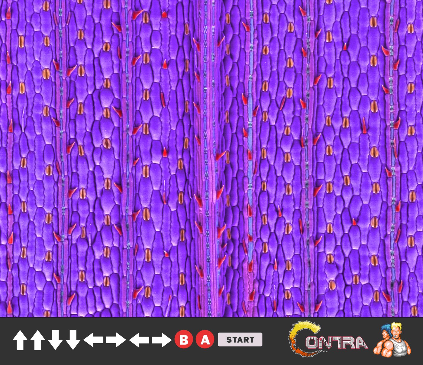
To accomplish this work, I’m really grateful that my friend Yu Yang from ZEISS China decided to share some of his brilliant SEM images and let me practice coloring. The sample is a Setaria Viridis leaf (狗尾草叶). Because it’s a fresh leaf he picked up outside his office, it was imaged using a coldstage and under variable pressure mode in a ZEISS EVO 15 SEM. Coloration was added in Adobe Photoshop.

Just like the other plant/grass in the Poaceae family, the leaf surface is full of small spikes which reminds me some of my childhood video gaming scenes. Of course, with Photoshop I have the flexibility to make something a bit wild.


Leave a comment